Imaging & Histology
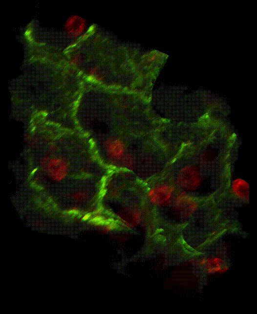
3D confocal microscope image of macrophages (red) in the mouse lung airways (green).
Image courtesy of Trudeau Institute Imaging & Histology Core Facility
The Imaging & Histology Core provides expert histology, immunohistochemistry, immunofluorescence, and confocal imaging support to our clients. With decades of experience, our in-house offering can be added to project designs to help visualize the impact of infectious disease and treatment protocols.
Available technical support may include existing standard operating protocols as well as development of new techniques and protocols as needed.
The following services are available for interested researchers. Please feel free to contact us regarding other requests.
Tissue Fixation
Paraffin & Cryogenic Embedding
Antigen Retrieval
Sample Decalcification
Protocol Optimization
Hematoxylin & Eosin (HE) Stain
Special Stains (by request)
Immunohistochemistry (IHC)
Light Microscopy
Immunofluorescent Microscopy
Confocal Microscopy